Of course, it’s hard to imagine that the microbes can be called beautiful. However, the photo competition of «Nikon Small World» presented in all its glory. The famous photo contest are micrographs taken with an incredible level of zoom. Photographers are able to find beauty in the most unexpected, but quite ordinary things, such as flea or a crystal of soy sauce. The below picture shows the beauty of pathogens, morbid cells and other elements of various diseases. Submitted photo took part in the competition «Nikon Small World» in 2010 and 2011.

Giant liposomes of pulmonary surfactant (40x) (Dr. Jorge Bernardino de la Serna, Center for Biomembrane Physics, Department of Biochemistry and Molecular Biology / Courtesy Nikon Small World)
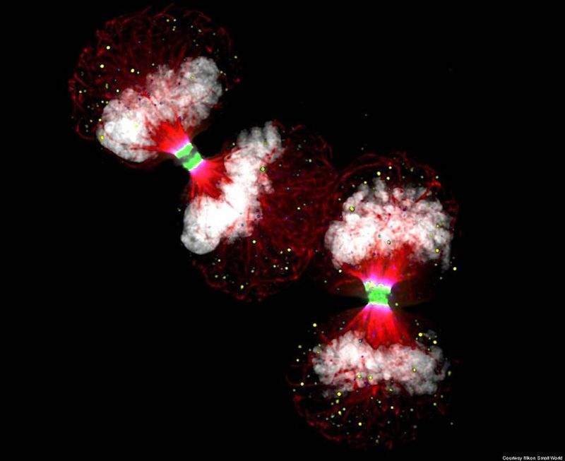
Telophase HeLa (cancer) cells expressing Aurora B-EGFP (green) (100X), Deconvolution (Dr. Paul D. Andrews / Courtesy Nikon Small World)

Anisakis pegreffi worm (40x) (J. Claire Hoving, Institute of Infectious Disease and Molecular Medicine, University Of Cape Town / Courtesy Nikon Small World)

Human Osteosarcoma cells (U2OS) (60x) (Dr. Ana Pasapera, NIH / Courtesy Nikon Small World)

HeLa (cancer) cells (300x) (Thomas Deerinck, National Center for Microscopy and Imaging Research / Courtesy Nikon Small World)

Three dimensional cell culture of breast cancer cells (MCF-7 cell line) (1000x) (Dr. Jônatas Bussador do Amaral, University of Sao Paulo / Courtesy Nikon Small World)

Nippostrongylus brasiliensis (rat nematode parasite) (560x) (Sinclair Stammers, Science Photo Library / Courtesy Nikon Small World)

Mast cell within collagen fibers (human eye – conjunctivitis) (7000x) (Donald Pottle, The Schepens Eye Research Institute / Courtesy Nikon Small World)
Beautiful Microbes by Nikon Small World (8 Photos)
previous post